Don’t Forget Your Back

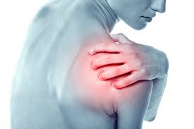
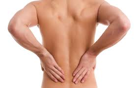
Most of us spend a large portion of the day hunched forward.  Think about when you are working on the computer, cooking, driving or even relaxing.  Your typical posture during these activities is head and neck forward, shoulders rounded forward, back curved and hips flexed.    When you remain in this position for an extended period of time, the muscles in the front of the body tend to shorten or contract, while the muscles in the back of the body tend to weaken.    This can be a set up for back, neck and shoulder problems and in addition, tends to make you look shorter, heavier and less confident.

Stretching before and after exercise as well as during the day is often overlooked in the rush to “get your workout in”.   Stretching however, has numerous benefits.   According to a study published in the Annals of Physical Medicine and Rehabilitation Medicine in September 2016, by I Fekhfekh, et al, dynamic muscle stretching of the knee musculature actually resulted in an increase strength gain in those muscles.  Interestingly, this study also found a decrease in the postural stability of the knee after stretching.    In English, it appears that stretching helps your to build strength, but may decrease the stability of the muscles you stretch at least temporarily.

In general, stretching helps to improve flexibility, joint range of motion, and usually is helpful for injury prevention.    Stretching helps by increasing blood flow to the muscles stretched.  This increased blood flow carries important nutrients to your muscles to allow for muscle growth and repair.  In addition, the increased blood flow helps to wash away muscle “waste” which leads to decreased soreness and inflammation of the muscle.

If allowed to remain in a contracted position for a prolonged period of time, our muscles will shorten.  For example if you spend a large portion of your day sitting in a chair or driving, the muscles in the front of the hips, your hip flexors will shorten.  These muscles have a direct effect on both your posture and your back health.


So what should you do?

It is important to stretch the muscles in the front of the body at least daily.

Exercises that focus on the hip flexors:

-Kneeling hip flexor stretch  – in the position of a lunge, allow your back knee to touch the floor and drive the hip of your front leg forward.  This will stretch the hip flexor of the knee on the floor

Pigeon stretch – extend your right leg straight back and bring the heel of your left leg underneath your right hip.  Then drive your hips forward towards the ground.  Stretch and repeat on the other side.

Yoga poses such as: low lunge, crescent lunge, upward facing dog, and revolving side angle pose


Stretches that focus on the chest muscles and prevent rounded shoulders:

Door Frame stretch- stand in a doorway or at the corner of a room with your arms up like you were going to stop traffic.  Line your arms up from the elbow to the hand with the doorframe or in the corners of the wall.   Step towards the door and you should feel a stretch in your chest (pectoralis “pec” muscles) and hold for 20-30 seconds.

Wall slides – stand with your back to the wall and keep your shoulders against the wall.  Walk forward 1-2 steps and slide your arms up and down the wall and squeeze your shoulder blades together.  You should feel a stretch in your upper chest muscles.

Shoulder squeeze stretch –  clasp your hands together behind your back and slowly try to lift your hands.  Squeeze your shoulder blades together to stretch out the chest wall.

Yoga poses include : bridge pose, camel pose, cobra pose and cow face pose


Stretches for your neck:

– Chin Tucks (Neck Retraction) – keep your shoulders back and your head in a neutral position (eyes facing forwards, chin level) slowly move your head backwards until you feel a slight stretch in the back of your neck.  Hold for 10-20 seconds and repeat.  If needed you can apply a gentle pressure on your chin with your fingertips to press your chin backwards and deepen the stretch.

Incorporating these exercises into your daily routine will help to prevent shortening of the muscles in the front of the body which leads to poor posture and often pain of the neck and back.  As always, if you are having pain which persists you should be evaluated by a certified health care professional to ensure that these exercises are safe for you.

This week we primarily discussed stretching exercises to prevent muscle contraction, and next week we will discuss the importance of strengthening the back muscles to further combat the issues of a hunched posture.

Common Causes of Knee Pain

The primary function of the knee is to bend and straighten the leg, to allow for a smooth gait during walking and running and to allow for jumping.    It is very common to develop knee pain in both new and seasoned exercisers.  Often the type of pain which develops is related to the type of exercise being done.

To understand knee pain, it is helpful to have a basic understanding of knee anatomy.

The knee joint can be divided into intra-articular and extra-articular compartments.

Intra-articular – structure inside the knee  joint

Extra-articular – everything else

The knee joint is where the surfaces of the leg bones meet and interact.  In order for the bones to move smoothly against each other and to provide additional stability, there is articular cartilage or cushioning shock absorber called the meniscus on both sides of the knee joint.  There are also many important ligaments which connect one bone to another and provide stability.  In addition, there are fluid filled sacs called bursae which allow for separation between solid tissues so that smooth movement can occur.

To help visualize the components of the knee, let’s break it down into 3 parts.

  • The anterior (front) of the knee
    •  Knee cap or patella
    •  Quadriceps tendon
  •  The medial aspect (side) of the knee  closest to the other knee
    • Medial meniscus (cushion)
    • Medial part of the joint capsule (the whole joint capsule encloses the entire knee)
    • Medial Ligaments  (provide stability for the knee and connect bone to bone)
  •  The Lateral aspect (outer) part of the knee
    • Lateral part of joint capsule
    • Lateral meniscus
    • Lateral ligaments

The knee is actually more complex then described above and does have the ability for rotation in addition to bending and straightening and other components not described above.  For the purpose of this article, we will focus on basic knee motions and components.

Now that we have a basic understand of the knee, let’s focus on what can go wrong with the knee.

In general, knee pain can be caused by:

  • Injuries due to trauma
  • Overuse injuries due to
    • Body misalignment
    • Muscle imbalances
  • Degenerative conditions
  • Injuries in the hips or feet

Patellofemoral syndrome (Anterior knee pain – front of the knee)

Common in new exerciser, especially runners and jumpers.  This is usually due to irritation between the patella (knee cap) and femur (thigh bone).  This pain can occur with increased jumping, squatting, running and climbing stairs.  Usually pain occurs if there is repeated stress on the knee or if there is a sudden significant increase in physical activity.   It is often treated with rest, ice, elevation and compression (RICE) and specific exercises to strengthen muscles surrounding the knee, and core.

Structural Injuries 

Knee injury can also result from structural changes or damage such as a dislocation, slippage or tears of the menisci or ligaments surrounding the knee.

Meniscal Injuries – The meniscus serves as a shock absorber for the joint.  Injuries of the meniscus can be the result of a twisting injury and can cause considerable pain, joint swelling, difficulty with stairs and the sensation of locking of the knee.  Occasionally the pain in the joint due to meniscal injuries will cause the brain to “shut off” the quadriceps at the knee causing the knee to “give way”.

Ligament Injuries – Ligament tears, such as a tear of the ACL (anterior cruciate ligament) can occur with a twisting injury and results in knee instability and may require surgical intervention depending on the degree of the tear.  It is common to sense a “pop” and immediate leg instability with this injury.    If surgery is used to repair the ACL, recovery is long and only low impact exercises are recommended initially to strengthen the muscles around the joint.  In contrast, PCL (posterior cruciate ligament) tears often can be treated non surgically with physical therapy and if indicated bracing.   When ligaments are torn or lax (loosened) it is common to have the sensation of poor balance or joint instability.

Tendon Injuries – Tendons are tissues that connect muscle to bone.  Typically, complete tendon ruptures are abrupt and result in the inability to move the joint.  The quadriceps tendon is quite large and a significant amount of degeneration must be present for it to rupture.  More often this injury is the result of a high force injury.   A completely torn tendon requires surgical intervention, but a partially torn one, will likely heal with immobilization followed by physical therapy.

Overuse injuries

Overuse injuries such as bursitis (inflammation of bursae), tendonitis (inflammation of the tendons), muscle strains, and Iliotibial band syndrome are usually the result of repetitive stress on a muscle, tendon or bursa.  Usually there is a component of body malalignment, muscle strength imbalance, improper shoe wear, or poor form which leads to the development of these conditions.  Treatment may include RICE (rest, ice, elevation, and compression) along with a detailed musculoskeletal exam to determine the etiology of the injury.  This will allow for a proper treatment plan to prevent recurrence.

Arthritis

Arthritis can be divided into Osteoarthritis (wear and tear), Rheumatoid (immune mediated), Gout (due to gout crystal build up) and Septic (due to infections).   Arthritis may be the result of poor diet or gluten sensitivity in some individuals.   While some forms of arthritis are genetic, diet modifications, weight loss, proper shoe wear and exercise may help to offset the severity of these conditions.

Rotator Cuff Tendonitis

The shoulder joint, or glenohumeral joint, is like a ball in a socket.  The shoulder joint is the interface between the humerus (arm bone), the scapula (shoulder blade) and the clavicle (collar bone).  Because the bony part of the socket of the shoulder is not deep enough to fully contain the top of the arm bone, there is a cartilage ring surrounding the bony joint to further contain and stabilize the arm.  The shoulder capsule is made up of fibrous tissue, helps to hold the arm bone in place inside the joint.   This capsule is fairly lax and allows for a good deal of movement in this joint which is both beneficial and detrimental.

The rotator cuff is a group of four muscles the help to move the arm in the shoulder socket but also serve to further stabilize the arm bone to allow for proper joint alignment and motion.  The 4 muscles in the rotator cuff joint are the supraspinatus, the infraspinatus, the teres minor and the subscapularis.  Each of these muscles have tendons, which serve to connect the muscles to the bone.  The most commonly injured muscle/tendon is usually the supraspinatus.  This is because the supraspinatus runs between two bony surfaces.  Depending on the anatomy of your shoulder, you might be at more risk for injury, especially if there is not much space between theses bony surfaces.  Over time, constant repetitive shoulder motion and friction causes micro-tears and fraying of these tendons.  High velocity injuries such as falls, car accidents or other trauma can cause also partial tears or rupture of these tendons and muscles as well.

Typically, rotator cuff tendonitis pain is brought on by specific movements of the shoulder joint.  Pain is usually located in the front or side of the arm and can radiate down to the elbow.  If the pain radiates further then the elbow, it is more likely your pain is due to a pinched nerve in the neck, rather than from your shoulder.

Rotator Cuff related pain includes (but is not limited to):

  • pain with overhead movements
  • pain with movements behind the back
  • pain with specific motions, but not all shoulder and arm movements
  • difficulty sleeping on the affected shoulder (this pain is also related to other shoulder pathology as well)
  • pain when raising or lowering the arm

Pain from the rotator cuff can be due to three different but often related issues. If the space between the bones is limited, when you raise your arm overhead, the rotator cuff can get pinched.  This is called impingement, which can be painful on its own.  Repetitive impingement can cause frays and tears of the tendons.    When the rotator cuff tendons get frayed this causes inflammation which leads to further development of pain.  Finally, the third reason for pain is shoulder bursitis.  The shoulder bursa is a fluid filled sac which allows for smooth motion between the tendons.  This sac or bursa can become inflamed and cause a significant amount of pain and point tenderness over the shoulder.

One of the reason for shoulder pain in improper body alignment.  In order for the rotator cuff to work optimally, the scapula or shoulder blade needs to move properly against the back of the chest wall.  Often times, during an exercise routine, you focus more on strengthening certain muscles but not others.  This can lead to muscle imbalance and improper alignment.   In addition, it is important to select the proper weights which are neither too heavy, no too light.  Your muscles should start to fatigue after 8-10 reps of heavy weight lifting but your form should not alter.  If you find that your form deteriorating, then you need to choose slightly lighter weights.

In my personal experience treating rotator cuff tendonitis, it seems that the body has a remarkable ability to heal itself if given the time to do so.  Unless you have specific anatomy which predisposes you to tears and impingement syndrome, usually the shoulder will recover within 6-12 months without much intervention.

That being said, there are interventions which may help with symptom management and prevention of recurrence.   Surgery might be required if you have a full thickness tear, a partial thickness tear and are a high level athlete, or anatomical issues which will not correct on their own.

Generally speaking, rotator cuff tendonitis is treated with rest, ice, and anti-inflammatory measures (either medications or dietary changes).  Sometimes, a cortisone injection into the shoulder is helpful if conservative treatments fail.  In addition, treatments such as PRP (platelet rich plasma) are used to help with regeneration of tissue.   After the initial rest phase, which should be short, as you don’t want to create a frozen shoulder, progressive shoulder motion exercises are recommended.  Once you regain shoulder motion, the next step is strengthening the muscles which fix the scapula (shoulder blade) to the chest wall.

Exercises such as:

  • Punches
  • Shoulder shrugs
  • Rows

are recommended next because proper strength and alignment of these muscles will help to fix the scapula motion with respect to the chest wall.  After you have re-established proper motion and increased the strength of these muscles (such as the rhomboids, latissimus dorsi, trapezius, serratus anterior, pectoralis muscles, etc), then you can progress to specific exercises of the rotator cuff muscle.

The reason you are often referred for physical therapy is that therapists are trained in the proper progression of these exercises and can instruct/monitor you for proper form and technique.  In addition, modalities such as taping, heat, ice, ultrasound, phonophoresis (using ultrasound to help medications penetrate into the shoulder joint, and iontophoresis (using electric current to help medications penetratethe shoulder joint may be effective, though the research studies are equivocal for some of these treatments.    Finally using a shoulder brace may help however, it is important to remove it often to prevent a frozen shoulder.

If you have shoulder issues, make sure to get examined by a medical professional so that you can begin the appropriate treatments and start on the road to recovery.

Exercising With Arthritis


We have all different levels of fitness. Some individuals have been exercising for years, while other individuals have never exercised a day in their lives.  We are all at different levels in terms of our cardiovascular fitness, our muscle strength and flexibility and our overall health.   It is quite important to remain active in spite of arthritis.  In fact, movement in people with arthritis is crucial to health and well-being.

Despite what we once thought, it is my experience that it is possible to modify the severity of certain types of arthritis.   In my mind, arthritis, chronic pain and other autoimmune diseases are the result of multiple factors.  All these factors such as genetics, environmental exposures, nutritional deficiencies, dietary influences, and gut health to name a few factors, all line up as if you were to “win” in the slots.  When this potent combination occurs in your body, havoc ensues.   The result is inflammation and chronic disease.   While you can’t change your genes per se, it is now thought that you can change or modify your gene expression.  In addition, you can certainly modify some of the other risk factors just mentioned to offset the severity of your debility.

In most cases, moderate exercise is essential to health and well being.  Proper, varied forms of exercise raises heart rate, raises endorphins, improves muscle strength, improves balance, improves flexibility and increases blood flow throughout the body including the muscles and joints.  In the case of arthritis, there is inflammation in the joint capsule and the bones themselves which leads to severe pain and often deterioration.     The nerve endings in the joint become super sensitive and pain can be unbearable.

Believe it or not, your choice of diet can directly impact your ability to exercise. When you consume an Anti-Inflammatory Diet, often you will notice you will able able to exercise with less pain is because this diet eliminates many known triggers for arthritis.


Some examples of arthritis triggers include:

  • Fried and processed foods
  • Thought to be due to compounds called AGE – (advanced glycation end products) These products are the result of heating, grilling, and frying foods.  The body utilizes pro-inflammatory molecules called cytokines in an effort to break them down and the result is excess inflammation in general as well as in the vascular system (blood vessels).
  • Sugary foods
  • Dairy
  • Alcohol
  • Foods/Oils with Omega-6 fatty acids – such as vegetable oil (safflower), corn oil, fried foods, certain nuts and seeds, mayonnaise


Some examples of beneficial foods include:

  • Foods containing high levels of Omega-3 Fatty acids – (ex. salmon, herring)
  • Avocados
  • Flax seed
  • Cherries and other berries (contain anthocyanins that are thought to be anti-inflammatory)
  • Leafy vegetables, broccoli – contain vitamin D
  • Green tea – has EGCG (epigallocatechin-3-gallate) a powerful antioxidant
  • Vitamin C containing foods (ex. papaya, brussel spouts, broccoli, citrus fruits)
  • Garlic, onions and leeks


It is very important to listen to your body.  Pay attention to the foods you are eating and the way you feel.  Often times, foods that are considered healthy may cause issues for you.  If you suffer from arthritis, try rotating your foods (i.e. do not eat the same food everyday).   If there are certain foods you think might bother you, wait 3 days in between eating them.  Then pay attention to the way you feel when you re-introduce those foods.  If you notice you have more difficulty with pain or decreased ability to exercise try eliminating the offending foods for a week or two.

Typically, it will take several weeks of following the diet closely before you see results with regard to decreased pain from inflammation.

When exercising with arthritis, it is important to modify your exercises as per trainer recommendations.  If exercises hurt, if possible, discuss with your trainers to find another way to exercise that muscle group.  In the beginning, there may be some exercises you simply cannot do.  What is important is that you are attempting to exercise.  With each passing week you will find that you can do a little more than the previous week.  Progress may be slow, with ups and downs.  As always, if despite closely following the diet, you are still having issues, it might be time to seek the advice of a health care professional.

Belly Fat


Body image and self-confidence can be greatly improved with physical activity and dietary changes, but arguably more important, are the numerous health benefits of exercise and proper nutrition.

One misconception about belly fat or abdominal fat is that you need to target only the abs to reduce belly fat. This is not exactly the case because the body is not very efficient at targeted fat burning.   Instead, fat burning exercises, such as aerobic exercises, burn fat fairly evenly throughout the body. Losing belly (abdominal) fat can best be attained by combining several techniques, rather then focusing on just abdominal exercises such as crunches and sit-ups.

It is important to take note of your body habitus or shape. By now, you have probably heard about the “apple” and the “pear” body shapes.

  • Pearshaped body: people carry fat around under the skin or “subcutaneously” in the hips, thighs and buttocks
  • Appleshaped body type carries a type of fat known as “visceral” fat, or fat stored in the abdominal cavity

The danger of this visceral fat in the abdomen is that it surrounds internal organs such as the liver, pancreas, kidneys, intestines, etc. Visceral fat cannot be pinched. You can be skinny and still have visceral fat.   Fat cells are not passive, but in fact, active cells. Visceral fat cells produce fatty acids and inflammatory cytokines (chemicals) that can drive or worsen existing inflammation (such as arthritis or cardiovascular disease).   In contrast, subcutaneous fat (fat you can pinch under your skin) produces beneficial hormones such as leptins that drive your sense of fullness.

There are many factors that determine the placement of fat on our bodies. Some are modifiable while others are not. Genetics definitely play a role on visceral fat, as does various hormone levels, but so to does your total calorie intake. In addition, the composition of your diet, including the amount of dietary protein, level of dietary sugar intake, and type of fat consumed in your diet, all have implications on fat storage and overall health.

Elevated visceral fat (belly fat) has been linked to:

  • Metabolic disturbances
  • Cardiovascular disease (strokes and heart attacks)
  • Diabetes
  • Cancers (such as breast)
  • Arthritis
  • Demenita, Anxiety, Depression
  • Sexual dysfunction

According to the American College of Sports Medicine, you are at significantly increased risk of cardiovascular events if you have a waist circumference of > 40 inches in males and > 35 inches in females.   There is a test you can do to determine if you are at an increased risk called the Waist-to-Hip circumference ratio. Using a tape measure, check the circumference of your waist at your narrowest point and measure the circumference at the widest point of your hips.   If your ratio of Waist/Hips is > 0.95 as a male or > 0.86 as a female you are at significantly increased risk for cardiovascular events, hypertension, and type 2 diabetes.

So what should you do?

Diet: this is the most important component of your strategy to reduce your abdominal fat and bloating.

  • Avoid foods which bloat you (different for different individuals)
  • Avoid chewing gum
  • Avoid simple sugars and processed carbs
  • Avoid alcohol – the liver will burn this instead of fat for energy
  • Avoid dehydration – Drink water
  • Avoid bubbly drinks as this can worsen abdominal gas and bloating
  • Avoid wheat/gluten
  • Chew your food until it is liquefied for better digestion
  • Consider adding a probiotic if o.k. with your Physician or Health Professional
  • Add 10 grams of soluble fiber to your diet –(1 cup of peas or 2 apples)
  • Avoid trans-fats which are stored as visceral fats

Exercise:

  • At least 30 minutes of moderate intensity exercise 5 days per week
    • High intensity interval training – helps burn visceral fat
  • Strength train 2-3 times per week to build muscle mass
  • Target all major muscle groups when strength training
  • Core exercises build abdominal muscle which will in turn burn fat all over body (but not locally around the abdomen)

Other:

  • 5-8 hours of sleep per night is optimal per study done by Wake Forest
  • Try to reduce stress – cortisol (a stress hormone) promotes visceral fat storage
  • Have your doctor check your hormone levels if you are worried – low testosterone in males and low estrogen in females can be associated with visceral fat storage

One caveat to this is that after a significant weight loss, you are sometimes left with excess skin. Excess skin is very different from abdominal fat. Unfortunately, at this time, I am unaware of any medically sound practices that reduce large quantities of excess skin aside from plastic surgery at this time.   That being said, the reduction of visceral fat and increase of muscle mass, due to proper diet and exercise, have profound, lasting effects on your health and well-being.

Hamstring Injuries


The hamstring muscles are actually a group of 3 muscles located in the back part of your upper leg. The hamstrings muscles originate from the pelvis (buttocks) and the femur (leg bone) on the backside of the leg and cross the knee joint to insert on the medial (inside) and lateral (outside) part of the leg just below the knee. The main jobs of the hamstrings are to extend (straighten) the hip and to flex (bend) the knee.   In addition, some of the hamstring muscles play a role in rotating the lower leg.

The most common thigh injury is a hamstring strain.   These injuries can often occur when the hip is flexed and the leg is extended, as in a single leg front kick. This is because 2 of the 3 muscles that make up the hamstring group cross over two joints (the hip and the knee).   These muscles are maximally stretched when the hip is bent and the knee is straight, and if movements are quick, the muscle can get strained or even tear.

Certain physical findings can increase your chances of getting a hamstring injury, such as:

  • Tight hamstring muscles – (inability to completely straighten your knee on standing)
  • An imbalance between the muscle strength in the upper leg (hamstrings and quadriceps)
  • Tightness of the quadriceps or hip flexor muscles (inability to completely stand up straight while the legs are straight — you will notice that you bend forward at the hips slightly if your legs are otherwise straight)
  • Insufficient warm up before exercising
  • Poor muscle coordination
  • Muscle fatigue
  • Muscle strength or flexibility imbalances

Usually a hamstring injury will present with pain on the backside of the upper leg, possibly with swelling, bruising, and an associated popping sensation at the time of injury.   If the hamstring muscle is fully torn, there may be an obvious mass which is actually the muscle contracting into a “ball”.

Treatment of hamstring injuries can be broken down into the acute phase that occurs immediately after injury and for the first 2-3 days. The sub-acute phase that occurs for several days to a few weeks after injury and the chronic phase that begins several weeks after injury.

Acute treatment of a hamstring injury generally follows the acronym PRICE.

Protection – In a severe hamstring strain or tear, an individual may require a crutch or a cane to protect the injured leg while walking.

Rest – for the first few days, remain off the injured leg as is possible, as this will enable the hamstring to begin healing.   The caveat is that stretching muscle begin shortly after injury because the muscle will scar down as it heals leading to further muscle tightness and predisposing you for another injury.

Ice– using a plastic bag filled with ice and water for 20 minutes 3 times a day is helpful to further reduce swelling. Heat is initially not recommended because heat increases the blood flow and is thought to worsen swelling. However, a few days after an acute injury heat can be helpful as heat increases blood flow to the injured area and helps with healing.

Compression – An ace wrap or compression dressing should be applied to the upper part of the thigh to prevent further swelling.

Elevation – Elevating the injured leg helps to further prevent swelling.

As mentioned above, hamstring injuries require both stretching and strengthening in order to recover. The main issue is that after a tear or strain, the muscles heal by scarring down. This scarring results in decreased flexibility of the muscle.

One big mistake athletes and weekend warriors make after sustaining a hamstring injury is to return too quickly to their prior level of activity. It is very important to only return to exercise after the pain has subsided. This is because if you return to exercise too early, you will change your biomechanics (i.e. alter your posture or positioning) to compensate for the pain. This altered position or step length while exercising can allow the muscle to shorten. When the muscle heals and contracts (shortens and scars down) you are then at increased risk for a repeat strain or tear.   Typically it takes at least 4-6 weeks for the muscle to properly heal.

It is helpful to begin stretching and strengthening exercises under the supervision of a qualified physical therapist so that you do not re-injure yourself and you can recover optimally. It is possible to regain or even surpass your pre-injury strength and flexibility with a proper treatment regimen.

PRP or Platelet Rich Plasma injections are sometimes recommended for assistance with tissue repair and quicker recovery time. However, an article published in 2015 from the Department of Sports Medicine at St Lucas Andreas Hospital in Amsterdam, reviewed the data for the treatment of hamstring injuries and found that lengthening exercises (stretching the hamstrings) provided the quickest return to play time, but did not affect re-injury rate. PRP injections did not improve outcome or re-injury rate when compared to controls. Therefore, at this time, it appears that a physical therapy regimen focusing on first stretching the hamstring muscles, then strengthening them (while maintaining improved flexibility) provides the best chance for recovery and return to exercise.   It was also suggested in this study that progressive agility training and trunk (core) strengthening and stability might reduce re- injury rates.

As always, if you suspect you might have a hamstring injury or other leg injury it is important to be evaluated by a trained health care professional. It is quite possible that you will be referred to a physical therapist for optimal treatment that includes a both a stretching and strengthening regimen. While initially your treatment will focus on the injured muscles, it will be important to analyze and treat your body mechanics to prevent repeated injury.

Carpal Tunnel Syndrome


One of the most common causes of hand and wrist pain is a condition called Carpal Tunnel Syndrome. This condition is usually the result of the median nerve getting trapped and compressed in the carpal tunnel as it courses towards the hand.

The carpal tunnel is a tunnel in which the bottom and sides of the tunnel are formed by the carpal or wrist bones and the top is formed by the transverse carpal ligament. This ligament or fibrous band connects the top two carpal bones. Inside the carpal tunnel is the median nerve as well as 9 of the flexor tendons for the fingers and thumb. The purpose of the carpal ligament is to stabilize the carpal bones and anchor some of the tendons for the hand muscles (finger flexors) to enable them to work correctly.

Carpal tunnel syndrome occurs due to compression of the median nerve running through this tunnel. The compression can be due to swelling of the tendons, inflammation of the tendons, overall fluid retention in the body (due to pregnancy, allergies, or other inflammatory conditions) and over-use injuries (such as typing).

Carpal tunnel syndrome can be classified as mild, moderate or severe.

Mild carpal tunnel presents as primarily sensory symptoms with numbness and tingling of the fingers and hand. As the median nerve gets more compressed the symptoms of carpal tunnel syndrome becomes more severe and the motor component of the nerve gets involved. When this happens, weakness and atrophy (muscle wasting) can be seen.

Common Symptoms of Carpal Tunnel Syndrome:

Mild:

  • Numbness, tingling or aching of the thumb, 2nd, 3rd, and part of the 4th fingers
  • Nighttime hand/wrist pain and numbness
  • Difficulty typing, or with fine finger movement involving the first 3-4 fingers
  • Numbness and tingling which worsens when the wrist/hand is bend at a right angle (either flexed forward or extended backwards)
  • Electric shock sensations radiating into the hand
  • Pain that radiates from the wrist upward towards the elbow

Moderate to Severe: (in addition to above symptoms)

  • Weakness in hand grip
  • Dropping objects from the hand
  • Muscle wasting in the palm of the hand, especially at the base of the thumb

For mild symptoms the use of a wrist splint at night will be helpful. These splints can be purchased over-the-counter at a pharmacy or medical supply store. The splint should have Velcro or adjustable type straps and should have some sort of support or rigid material on the underside of your wrist and palm, that will prevent your wrist from bending forward too much. The splint should be worn at night to prevent inadvertent wrist bending, and can be worn during the day if symptoms are continuously present.

Certain exercises may exacerbate carpal tunnel syndrome such as push-ups, planks, and low jacks. Any exercise which puts pressure through the wrist while it is at a 90 degree angle from the arm can further compress the median nerve in the carpal tunnel. Helpful exercise modifications include doing push-ups on your fists and keeping your wrists straight, or doing the push-up on an incline (with your head higher than your feet) so you are putting less weight through your wrists.

In addition, activity modification may be indicated if the above interventions are not helpful. If cleared by your doctor, adding a small amount of Vitamin B6 (<200mg/day) is beneficial for improvement of nerve function. Vitamin B6 acts a a diuretic (decreases swelling), helps the body to make neurotransmitters and helps with the function of the immune system.

Foods which contain B6 include:

  • Wild caught tuna
  • Bananas
  • Salmon
  • Grass-fed beef
  • Chicken breast
  • Spinach

If dietary, activity, and exercise modification are not successful in treating your carpal tunnel syndrome in conjunction with the use of a wrist splint and occasional anti-inflammatory medications, it is important to be evaluated by a health care professional. In addition, if your symptoms progress, are not localized to your first three fingers, or are associated with any other symptoms, make sure to get checked to rule out other causes of hand/finger numbness.

If conservative treatments fail, other interventions such as physical therapy, corticosteroid injections, prolotherapy, platelet rich plasma injections, or surgical release may be indicated.

Plantar Fasciitis

Plantar fasciitis is a common cause of foot and heel pain. Typically this pain is located on the bottom of the foot just in front of the heel pad on the inner or middle side of the foot. The plantar fascia is a thick tendon that connects the heel bones to the toes. This tendon serves as a shock absorber for the foot and helps in supporting the arch of the foot. Overuse or excessive stress on this tendon can cause small tears that lead to inflammation and significant pain.


Some common risk factors for plantar fasciitis include:

  • Obesity
  • Poor foot mechanics
  • Improper shoe wear
  • Long distance running
  • Jumping exercises
  • Prolonged standing

Exercise regimens that consists of a fair amount of jumping or plyometric exercises are very good for cardiac conditioning and development of muscle power. Generally speaking the heart muscle gets conditioned faster then skeletal muscle. Therefore, your skeletal muscles will fatigue more quickly and your form will break down before you get exhausted due to deconditioning from the heart.

There are muscles on both sides of the foot and leg that help to support the foot and ankle. The peroneus longus and peroneus brevis insert on the outermost or 5th metatarsal (mid bones of the foot) and the tibialis anterior inserts on the inner or 1st metatarsal bone. When these muscles are strong and conditioned, they help with stabilizing lateral (side-to-side) foot motion and control the rate of movement in the foot during jumping exercises. If however, these muscles fatigue quickly or are not yet strong, the foot and ankle has more mobility and collapse in the arch, which in turn can put more strain on the plantar fascia.

This is one of the reasons that plantar fasciitis typically develops in an exerciser after several weeks or months of exercise. It generally occurs when the aerobic or cardiac conditioning out paces the skeletal muscle conditioning. In other words, your heart endurance allows you to jump or run for longer periods of time, but your leg muscles are not conditioned enough to control your ankle motion. If in addition to decreased muscle strength, you are carrying around extra weight, are wearing improper shoes, have poor foot mechanics or are increasing the amount of time or the intensity of your jumping, you are a set up for plantar fasciitis.

So what should you do?

It is important to know what type of foot you have and have the proper shoe wear before embarking on exercise and lifestyle changes. There are several very good running stores and even some online stores which will question you about your feet and recommend proper shoe wear. Podiatrist or Sports Medicine/Rehabilitation Medicine physicians are also useful for providing recommendations and treatment plans. It is important to note that not one type of sneaker is good for everyone. Sometimes due to foot or even hip and knee mechanics you will need an orthotic insert to place in your shoe.

In general, a helpful guideline for shoe wear and orthotic use is to fix what is too flexible (i.e put in arch support in a very flat foot or over-pronating foot) and to accommodate foot deformities that are fixed (i.e. relief or padding around bony deformities such as bunions). A common mistake is to over correct too much (using too rigid or too high an arch support). This can lead to other issues higher up the kinetic chain such as knee, hip or back issues.

Plantar Fasciitis is typically worst in the morning just after getting out of bed. Pain is usually severe with initial weight bearing and then subsides throughout the day with walking and weight-bearing. Pain can also be severe after prolonged sitting or immobility. It is often helpful to wear nighttime splints to keep the foot/Achilles tendon stretched out overnight. This will decrease the initial severe morning pain because the fascia will have more mobility and will not be able to scar down overnight.

Aside from using proper shoe wear and night time splints, occasional use of anti-inflammatory medications such as Motrin will be helpful. Adding anti-inflammatory foods such as turmeric, (curcumin), boswellia, and Omega-3 fatty acids in addition to decreasing intake of Omega-6 fatty acids may also be helpful. Icing can be helpful as well. Sometimes, despite the above interventions, you will might need a cortisone injection into the plantar fascia to decrease inflammation and pain.

During the acute inflammatory phase of plantar fasciitis, it is important to limit the activities such as high impact jumping and running that can aggravate the condition. Modify your exercise to limit jumping until pain resolves during activity and upon wakening. As pain decreases is it imperative to resume jumping/high-impact exercises slowly over a period of weeks to even months to prevent recurrence. As with all my recommendations, if you have persistent heel pain that is refractory it is important to seek the advice of a medical professional to make sure other modifications or treatments are indicated.